Muscle Anatomy
Striated Muscle
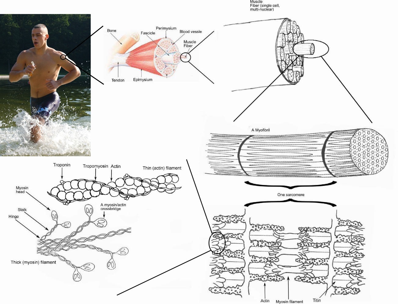
Muscles are composed of bundles of muscle fibres wrapped in a fasicle which run longitudinally along the muscle length.
A muscle fibre is a multi-nucleated single cell. This multi-nucleation is due to how the muscles are formed during embryonic development: several stem cells (myoblasts) fuse to produce each muscle fibre.
The smooth endoplasmic reticulum of the muscle fibre is known as sarcoplasmic reticulum (SR).
- During rest, Ca2+ ions are continually actively transported (ATP-dependent) into the SR
The plasma membrane of a muscle fibre (sarcolemma) has tubular infoldings (T tubules) which go through the cell and out the otherside.
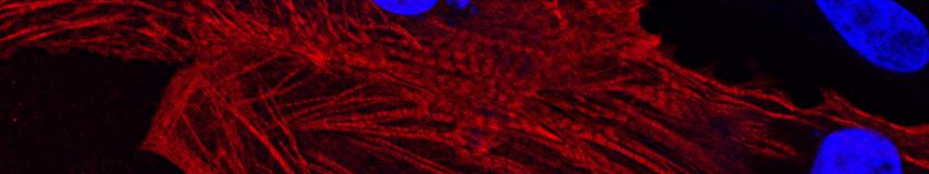
The cytoplasm of the muscle fibre is called the sarcoplasm. This contains long protein bundles of myofibrils.
The striations in muscle are due to the presence of sarcomeres in the myofibrils.
Sarcomeres
These are the contractile units of the myofibrils.
They are composed of thick filaments and thin filaments.
Sarcomeres are separated into different bands:
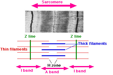
- Dark A bands alternate with lighter I bands. The A band is dark due to the presence of thick filament. The I band is lighter as there is only thin filament present.
- The darkest part of the A band is from the overlap of thick and thin filaments in this region.
- The lighter region in the middle of the A band is the H band: thin filaments do not reach here.
- The dark line running down the middle of the I band is the Z disc/line which is made up of the elastic filament titin.
- It's to this region that the thin filament anchors to.
- This is also the border of the sarcomere.
Myofilaments
Thick filaments:
- 15nm in diameter
- Made up of several hundred molecules of myosin
- One molecule is composed of 2-intertwining-polypeptide tail and a double globular head that sticks out at an angle
- The myosin heads project outward in a spiralling manner
- These can bind ATP and hydrolyse it to ADP + Pi
Thin filaments:
- 7nm in diameter
- Composed of 2 intertwined strands (F actin) of "beads" of actin (G actin)
- Each G actin has an active site that can bind to a myosin head
- There are also 40-60 molecules of tropomyosin present
- These block the active sites of 6/7 G actins, preventing the binding to myosin
- On each tropomyosin molecule, there is also a smaller calcium-binding protein (troponin) bound to it
Elastic filaments:
- 1nm in diameter
- Made up of titin
- Anchor the thin filaments to the Z line